Blog

September 22, 2017

The Plastics of Capri

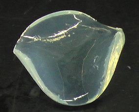
vase.jpgVitrum Flexile, or flexible glass. Although lost in legend, three different authors provided an account of a glass that could be dented and then repaired in Rome’s first century. According to the Corning Museum of Glass, Petronius (who died in 63 A.D.) told of a drinking vessel presented to Emperor Tiberius (reign 14-37 A.D.). The vessel was thrown to the ground, and though dented, the glassmaker was able to remove the dent with a hammer.

Pliny told a similar story in his encyclopedia, completed around 78 A.D., although his story goes on to say that the glassmaker's workshop was destroyed so that the value of copper, silver and gold would not be affected by the vitrum flexile. 

Dio Cassius provides the third story, in which an architect, who fell out of favor with Tiberius, provided the aforementioned demonstration of denting a glass vessel, then repairing it with his hands.

Unfortunately, no other accounts of vitrum flexile exist beyond these stories. As polymer scientists, we naturally assume that the vitrum flexile was not glass, but rather an early form of plastic, given its mechanical behavior and appearance. Perhaps it was formed of a natural resin, or from a crude polymerization. Ethylene gas was known to be in the Mediterranean around this time (the suspected source of the Oracle of Delphi’s visions). Did a resourceful Greek manage to perform some early free radical polymerization?

Posted by CatherineCerasuolo
0 Comments
September 20, 2017

It's a Plastic Sort of World

plastic capgs.jpg 

Provocatively Titled Study Explores “All Plastics Ever Made”

A recent study by Geyer et al published in Science Advances takes a wide-ranging look at the production and fate of all plastic produced to date, based on data collated from a range of market research and consulting groups.

Some key observations from the authors’ study are as follows:

The total amount of polymer resin produced from 1950-2015 is approximately 7800 Mt. Production growth has been accelerating with a compound annual growth rate of 8.4%—for perspective, half of the 7800 Mt has been produced in last 13 years!

Assuming an average pellet mass of 25 mg, the total amount of resin produced through 2015 is the equivalent of 3.12 * 1017 pellets.

Plasticizers, fillers, and flame retardants account for approximately 75% of all produced additives between 2000-2014. Antioxidants and heat stabilizers account for approximately 11% of additives produced.

Industrial Use of Plastics 

The largest industrial use sector for plastics is packaging. In fact, 42% of all produced nonfiber plastics are used for packaging purposes. The second largest use sector is construction, which makes use of 69% of all PVC produced.

GlobalPrimaryPlasticsProductionaccordingtoindustrialsectorusecropped.pngfig. S1. Global primary plastics production (in million metric tons) according to industrial use sector from 1950 to 2015.  [1]

The most commonly produced plastics are polyethylene and polypropylene, which make up 57.3% of all polymer resins produced from 2002-2014.

GlobalPrimaryPlasticsProductionByPolymerType.PNG

              fig. S2. Global primary plastics production (in million metric tons) according to polymer type from 1950 to 2015. [2]

Where Is All That Plastic Now? 

It is estimated that 30% of all plastics ever made are currently in use—the rest having been disposed in one way or another. It is estimated that 12% of all plastics disposed of have been incinerated, and only 9% have been recycled. The remainder are in landfills or the natural environment. Note that none of the commonly used plastics are naturally biodegradable; the vast majority are derived from petroleum hydrocarbons.

In addition, there is an increased attention of to the ultimate fate of polymer fibers (such as those used in textile/clothing applications). Environmental accumulation of polymer fibers is in many respects analogous to recent concerns about the deliberate addition of polymer microparticles to consumer products—recall the Microbead-Free Waters Act of 2015 which banned the manufacture and sale of rinse-off cosmetics containing polymer microbeads.

How CPG Can Work With You

At Cambridge Polymer Group, our expertise and analytical capabilities intersect with the topics raised by such studies in multiple ways. We can assist your team in:

  1. Material selection and screening
  2. Resin equivalency studies
  3. Identification of unknown polymers
  4. Characterization of material stability and polymer degradation
  5. Identification/quantitation of chemical degradation products
  6. Impact of additive packages on polymer and device functional properties

CPG is an ISO 9001 certified, 17025 accredited contract R&D and analytical testing lab. Contact us to see how we can work with you.


Posted by CatherineCerasuolo
0 Comments
September 15, 2017

Additive Manufacturing of Medical Devices

Additive Manufacturing.jpg

Most traditional machined components involve starting with a standard block of material, and then machining away material to form the final finished device. The advent of 3D printing systems has allowed the opposite approach to machining, where material is sequencing added to a part, in effect growing it from raw material with little scrap material generated, hence the term ‘additive manufacturing’. 3D printing allows generation of more complex geometries, such as porous structures, fully-enclosed hollow features, and finer structure than may be achievable with more conventional milling, lathing, or molding processes. Additionally, 3D printing provides the opportunity for patient-specific implants derived from X-rays or MRIs of the patient’s anatomy.

A variety of materials have been used for 3D printed medical devices, including titanium powder and its alloys, calcium phosphate powder, thermoplastics such as polyether ketone ketone, and UV or light-curable resins. Since 2013, the FDA has been clearing medical devices and drugs made from 3D printing technologies through either the 510(k) process or the emergency need process. In the 510(k) process for a device, a device is cleared by the FDA if it is shown to be as safe and effective as a legally marketed predicate device to which it is substantially equivalent (same intended uses, comparable design and materials). The same tests conducted on machined or molded devices required for clearance, such as biocompatibility, leachables/extractables, mechanical behavior, cleanliness, and functional performance, all apply to 3D printed devices, and currently no additional regulation specific to 3D manufacturing has been established. As of 2016, more than 85 devices manufactured using 3D manufacturing processes have been cleared by the FDA.

3D Printed Medical Devices

Some examples of 3D printed devices that were cleared are shown below, spanning a variety of medical application areas.

3dmeddev.jpg

2013

  • Oxford Performance Materials received 510(k) clearance for a 3D printed cranial-facial device made from laser sintering polyether ketone ketone (PEKK), granted in 2013.

2015

  • DENTCA, a 3D printed denture system, received 510(k) clearance in July of 2015. DENTACA is made from a light-cured resin, and underwent biocompatibility testing per ISO 10993, in addition to human mouth tissue sensitivity contact.
  • The first 3D printed drug to be cleared by the FDA was Aprecia Pharmaceuticals Co.’s epilepsy drug, Spritam® (levetiracetam) tablets in 2015. Spritam is for treating seizures in patients with epilepsy, and is printed using a method called ZipDose technology, which produces a porous pill that dissolves rapidly with liquid.
  • MedShape received 510(k) clearance on a Ti6Al4V 3D printed bunion correction system in 2015.

2016

  • K2M received clearance for titanium printed porous spinal fusion devices made with laser fusion, designed to have surface roughness with feature sizes of 3-5 microns to encourage bone on-growth.
  • Stryker received 510(k) clearance for a titanium printed lumbar cage to treat degenerative disk disease.
  • ZB’s reconstructive wedges made from OsseoTi (titanium powder) for ankle fusion received clearance. 
  • Additive Orthopedics cleared for titanium 3D printed devices for foot applications.  
  • Bioarchitects received clearance for 3D printed titanium craniofacial implants.

2017

  • OssDsign received 510(k) clearance for cranial plates 3D printed with a proprietary calcium phosphate composite reinforced with titanium in 2017. 

The Future of Additive Manufacturing 

3D printing of drugs could allow hospitals to customize the API dose in each tablet for individual patients, ushering in a new era of personalized medicine. Additive manufacturing may also make medication easier to swallow, both for adults due to improvements in swallow-ability and dissolution, and for children, in that the drug can be made in kid-friendly shapes. At this point, conventional large scale manufacturing continues to be more financially efficient for large quantities of medication, and FDA clearance of 3D manufactured drugs is not easy to obtain.

Additive manufacturing is becoming a more viable alternative manufacturing process for medical devices, since it offers solutions to designs that are unprocessable by conventional manufacturing approaches, readily allows custom device manufacturing, and may present options for on-demand manufacturing of implants. In addition to the standard tests for new medical devices, the areas that currently need addressing for additive manufacturing include process validation procedures and qualification of raw materials (new and potentially used).

Posted by CatherineCerasuolo
0 Comments
September 13, 2017

Shrimp Vision

Polymer scientists often use polarized light to optically examine features in polymers, such as crystallinity and residual stress, through the phenomenon of birefringence. White light, such as that coming from the sun and incandescent light bulbs, is comprised of multiple wavelengths of light traveling in multiple planes. When white light passes through a polarizer (which is a type of linear optical filter that only permits waves oriented in the same direction of the linear direction of the polarizer to pass through, and blocks all others, as shown in Figure 1), the randomly oriented light becomes plane-polarized in a single optical plane. 

polarized light.pngFigure 1: Schematic of randomly oriented white light passing through a polarizer, becoming plane-polarized light with the principle axis indicated by the blue arrow.

A similar phenomenon occurs when white light is reflected off of a flat surface, such as a body of water or a metal or glass surface. You may have experienced this phenomenon when wearing polarized sunglasses while looking at the ocean or your smart phone. In some orientations of the glasses relative to the ocean or phone, the light intensity is greatly diminished (and you may not be able to see the images on your phone) because the polarizer in your glasses is blocking a percentage of the plane-polarized light reflecting off the surface of the object you are looking at. By rotating your head in a clockwise fashion, the light intensity will decrease or increase as angle of the plane-polarized light moves towards or away from 90º from the principle angle of your glasses.  More details on the math associated with light polarization can be found on our birefringence application note.

The use of polarization of light in nature is commonplace. It has been known for decades that insects (bees, ants, beetles) use the atmospheric polarization of light[1] to navigate to and from their home bases.[2] Whereas these insects do not have traditional polarizers, they do have specific arrangements of photoreceptors that maximize the absorption of polarized light. Using polarized light aids in the sky and in reflected light off the ground and off plants, the insects can ably navigate. In most cases, the photoreceptor systems are fixed relative to the animal’s eyes. However, it was recently discovered that the mantis shrimp is able to rotate its eyes independently, effectively adjusting the principle orientation of its ‘polarizer’ to maximize the signal of incoming polarized light (see Figure 2). What do they do with this information? Identify food sources, for one thing. Many of the jelly-fish and multi-cell organisms in the ocean are nearly transparent to the naked eye. However, to a creature tuned into polarized light, these amorphous organisms light up against the dark ocean back ground as the plane polarized sunlight passes through their bodies, making them easy targets for hunting by the shrimp (see Figure 3). 

Rotational Degrees of Freedom.jpgFigure 2: Rotational degrees of freedom in Odontodactylus cultrifer.[3] 

Transparent.jpg

Figure 3: Transparent organisms made visible with polarized light.[4]

But the shrimp have a secondary use for polarized light, namely communication. Scientists have noticed distinct color patterns on shrimp that are only visible when observed through a polarization filter. As shown in Figure 4, the male shrimp has a distinct red pattern visible when the polarizer is oriented horizontally. The female (on the right) does not have this pattern, suggesting that the pattern may be useful for shrimp courtship.

MaleFemaleMantisShrimps.jpg

 Figure 4: Male (left) and female (right) mantis shrimps, showing a clear difference in polarizing signals (the arrows show the orientation of the camera polarizing filter). This method of communication  may be important for mating.[5]

Let’s hope that shrimp continue to use this optical power for food and mating, so that they don’t push us polymer scientists out of our jobs.

PolymerScientistShrimp.png


[1] The unpolarized light from the sun is scattered by water molecules, gas molecules, and particles in our atmosphere, resulting in localized polarization in regions of the sky.

[2] Wehner, “Polarized-light navigation by insects,” Scientific American, 106 (1976).

[3] From Daly et al., “Dynamic polarization vision in mantis shrimp,” Nature Communications 7 (2016). https://www.nature.com/articles/ncomms12140

Posted by CatherineCerasuolo
0 Comments
September 1, 2017

You Don’t Look Your Age: Accelerated Aging of Paper

oldbooks.jpgAt Cambridge Polymer Group, we help many of our clients evaluate their materials or device by applying accelerated aging techniques to accelerate material degradation, either for shelf life estimation or as part of an evaluation of material stability. Such testing may be performed following standard procedures like ASTM F1980, ASTM F2003, as well as custom aging studies which expose the material to specific environments which a particular product or material is expected to experience during end use. Selection of accelerated aging conditions requires careful consideration of the device composition, its materials’ properties, storage conditions, the end use environment, and the assumptions implicit in the accelerated aging calculations. As such we are always interested in test methodologies and case studies of material aging (whether accelerated or real time).

Accidental Accelerated Aging of Paper

Cellulose.png

Consider the storage of paper. Paper is composed of principally cellulose fibers, which are a naturally derived polymer—a polysaccharide with D-glucose as the repeat unit. Anyone who has dug in the far back corner of the university library basement stacks or through a box of books in Grandma’s stuffy attic knows that paper is subject to yellowing, degradation, and that “old book smell”. Naturally, for important documents, steps are implemented to preserve the documents and slow the degradation process. But such mitigating practices have in some cases been counterproductive.

Lamination 

Starting in the 1950s, South Carolina's state archives and history department laminated many thousands of documents for the purpose of preserving them. Seems ok, right? Sealing the paper away from the outside environment should prevent moisture and oxygen from causing damage. Except the opposite result has been observed after many years—the laminated documents are observed to yellow and even produce the scent of vinegar. Something’s wrong!

Sheet Proximity

Similarly, researchers at the Library of Congress performing accelerated aging studies tested cases where individual sheets of paper were left exposed to the atmosphere or placed in stacks containing many sheets in close proximity. Intuitively, one may expect that the sheets more exposed to the environment would age faster due to their greater exposed surface area. The opposite result is observed—the Library of Congress study found that stacks of paper were the ones aging more rapidly. What’s going on?

The Importance of Material Chemistry

The intuitive reasoning above ignores an important potential source of degradation; the paper itself and its protective materials - whether lamination or adjacent paper sheets. Researchers at the Library of Congress have found that acids (even trace acids in “acid free” paper) from within the material stimulate acid hydrolysis of the cellulose, a byproduct of which is additional acids which cause more hydrolysis and degradation. As a result, even acid free or pH neutral papers have been observed to become increasingly acidic as they age. When such paper is laminated or stacked among adjacent sheets, such acids are allowed to build up and, counterproductively, accelerate damage to the document. It should also be noted that such acid and volatile organic compound (VOC) byproducts of the paper degradation are responsible for that “old book smell” in the university stacks (so perhaps don’t breathe too deep).

The above situations are excellent case studies on the importance of carefully considering the chemistry of the underlying material prior to making design decisions on material storage or experimental decisions for accelerated aging studies (which may yield invalid results if founded on faulty assumptions).

Do you need assistance with accelerated aging, experimental design, stability testing, or volatile organic compound analysis? Contact Cambridge Polymer Group to see how we may be able to assist your team. 

Posted by CatherineCerasuolo
0 Comments
August 14, 2017

Cambridge Polymer Awarded $225K SBIR Grant from NIH

CPG research team to develop injectable hydrogel technology for eye tamponade

eye.jpg

Cambridge Polymer Group Inc has been awarded a $225,000 Phase 1 Small Business Innovation Research (SBIR) grant by the National Institutes of Health (NIH) to develop an injectable hydrogel for use as an eye tamponade.

Retinal detachment is a common eye injury that often causes vision loss in patients. The surgical procedure requires the temporary removal of the vitreous humor, the clear gel in the eye, prior to reattachment of the retina to the back of the eye. Following the operation, the retina needs to remain in contact with the back of the eye during the healing process, or it may become detached again. Surgeons currently achieve this process by either using a silicone oil, which distorts vision and requires a second surgery to remove, or with a gas bubble, which requires inconvenient head placement for the patient.

As an alternative, Cambridge Polymer Group’s team of hydrogel scientists are developing a hydrogel that can be injected in as liquid through a fine needle or cannula, and then gels in the eye. This hydrogel will assist in keeping the retina attached to the back wall of the eye, and will then degrade over time, obviating the need for a second surgery. The hydrogel should also allow vision through the eye while it is in place, resulting in greater patient mobility and compliance.

Cambridge Polymer Group’s Vice President of Research, Dr. Gavin Braithwaite, is the principal investigator on the research grant. “We are pleased with the opportunity the NIH SBIR program has given us to move our patent-pending technology from the laboratory bench into pre-clinical studies,” stated Dr. Braithwaite. 

For more info, see the press release.

Posted by CatherineCerasuolo
0 Comments
August 8, 2017

Hyaluronic Acid: A Question of Size

HA2.jpg

Figure 1: Top right—repeat unit of hyaluronic acid, a material particularly concentrated in (among other tissues) chicken combs (left). Bottom right—representative triplicate determinations of three samples of hyaluronic acid of distinct molecular weights.

Chemical Properties of Hyaluronic Acid

Hyaluronic acid (HA) is a naturally synthesized highly linear polysaccharide found in a variety of tissues including the joint space, vitreous humor, connective tissue, synovial fluid, and even chicken combs (Fig 1 left). Naturally synthesized HA is typically of a very high molecular weight well over 1,000,000 g/mol; it is extremely hydrophilic, highly lubricious, undergoes degradation in response to naturally occurring hyaluronidase enzymes, and binds to a variety of biological receptors. Such properties have driven interest in the use of HA in a variety of applications such as for the treatment of osteoarthritis knee pain, drug delivery/targeting, and tissue engineering.                                                        

In a physiological solution, the HA molecule is understood to exist in a twisted and entangled ribbon structure evolving from a combination of its repeat unit structure, internal hydrogen bonding effects, ionic interactions with the solvent, and its highly linear structure. These properties, in conjunction with the molecular weight of HA, have been shown to strongly affect its functional rheological properties, as has been reported by CPG scientists in the past.[1]

The accurate assessment of HA’s molecular weight therefore is an invaluable tool for applications such as material characterization, stability assessment, delivery behavior, and quality control. However, the same properties which lend to its unique properties also make molecular weight analysis of the material extremely challenging.

Molecular Weight Determination of HA

Polymer molecular weight is commonly determined by gel permeation chromatography (GPC; also known as size exclusion chromatography or SEC). However, due to the polymer’s extremely high intrinsic viscosity, large hydrodynamic volume, slow reptation rate, highly linear structure, and propensity for entanglement effects and shear degradation, analytical conditions must be chosen extremely carefully.

Some key analytical process variables which may dramatically impact measured molecular weights include sample concentration, dissolution time/temperature/agitation, solution handling, sample filtration (or lack thereof), mobile phase selection, injection volumes, flow rate, column selection, and calibration methodology. No conventional molecular weight calibration standards exist which may be used for HA analysis, and as a result, absolute molecular weight analysis by light scattering or triple detection is most commonly performed instead for accurate MW measurement.

CPG has validated an in-house method for the determination of hyaluronic acid molecular weight distributions by triple detection GPC (Fig 1, lower right). Contact us for additional information on this methodology and to see how CPG can work with you. CPG is an ISO 17025 accredited contract research and analytical testing lab based out of Boston, MA.


[1] Gavin J. C. Braithwaite, Michael J. Daley & David Toledo-Velasquez (2016) Rheological and molecular weight comparisons of approved hyaluronic acid products – preliminary standards for establishing class III medical device equivalence, Journal of Biomaterials Science, Polymer Edition, 27:3, 235-246,  DOI:10.1080/09205063.2015.1119035


Posted by MaryOsward
0 Comments